Mobility disappears slowly. A missed step here, a stiff morning there, and suddenly the ankle that used to glide feels like a stubborn hinge. I see this arc weekly, from runners after a sprain to people who never fully recovered normal motion after bunion surgery or a fracture. The good news is that feet and ankles respond to thoughtful, consistent work. With the right mix of assessment, targeted exercises, and evidence-driven therapies, the joint can recover glide, the soft tissue can regain length, and the brain can learn trust again.
This is a clinician’s guide built for real life. It prioritizes what reliably improves foot and ankle motion across common conditions, when to push and when to pause, and how a foot and ankle mobility specialist structures care. If you are already working with a foot and ankle doctor or physical therapist, compare notes. If you are not, use this as a roadmap and a nudge to find a qualified foot and ankle care expert who can tailor the plan to your anatomy and goals.
Motion that matters: dorsiflexion, plantarflexion, inversion, and eversion
Before therapies, we need a shared language. The ankle is primarily a hinge where the tibia and talus meet, with a subtalar joint beneath that handles side-to-side motion. The motions you feel and measure:
- Dorsiflexion, pulling the toes toward the shin. You need this to squat, descend stairs, and walk efficiently. A short calf or stiff joint often limits it. Plantarflexion, pointing the toes down. Essential for pushing off, sprinting, and balance recovery. Inversion and eversion, rolling the sole inward and outward at the subtalar joint. Critical for adapting to uneven ground and distributing load.
In practice, lack of dorsiflexion is the most common mobility roadblock I treat. Runners with Achilles pain, hikers with recurring sprains, patients with midfoot collapse, even people with knee issues often trace back to tight calves or a talocrural joint that stopped gliding.
How a foot and ankle mobility specialist evaluates stiffness
A skilled foot and ankle physician, whether an orthopedic surgeon or a podiatric specialist, starts with specifics. Where is motion stuck, and why?
I rely on several quick measures that guide both diagnosis and the exercise plan:
- Weight-bearing lunge test for dorsiflexion. Place your foot perpendicular to a wall, knee tracks over the second toe, heel stays down. Slide the foot back until the knee barely taps the wall without the heel lifting. Many healthy ankles reach 8 to 12 centimeters. Less than 5 often correlates with stiff joint mechanics or tight soleus. Silfverskiöld test to separate gastrocnemius from soleus tightness. If dorsiflexion improves with a bent knee, the gastrocnemius is the limiter. If it doesn’t, the soleus and joint capsule likely need work. Subtalar mobility and midfoot spring. Gentle inversion and eversion of the heel and a midfoot “spring” test indicate whether the restriction lives below the ankle or in the forefoot. Talocrural joint glide. A posterior glide of the talus should feel smooth. A “block” sensation often signals capsular stiffness that responds to mobilization. Gait and single-leg control. Watch stance phase, heel rise timing, and arch behavior. If the foot collapses or the heel won’t evert, the peroneals and posterior tibialis may be underperforming.
A foot and ankle joint specialist will pair this with imaging only when red flags arise, like trauma, persistent swelling, locking, suspected osteochondral lesions, or suspected stress fractures. X-rays show bony alignment and arthritis patterns. Ultrasound clarifies tendon involvement. MRI is reserved for complex cases, surgical planning, or failed conservative care.
The difference between joint stiffness and muscle tightness
People often stretch endlessly without moving the needle because they are chasing the wrong limiter. Here is the rule of thumb I share in clinic:
- If you feel a deep pinch in the front of the ankle when bending the knee over the foot, joint mechanics need attention. Posterior talar glide, heel cord soft tissue work, and closed-chain mobilizations help. If you feel a calf stretch that eases with the knee bent or straight in specific ways, the gastrocnemius and soleus are the primary players. Eccentric loading and long-duration stretching work well. If the arch collapses or the foot rolls in when you load it, mobility might be fine but control is poor. Foot-intrinsic training and tendon conditioning become the priority. If stiffness shows up only in the morning and eases as you move, think plantar fascia loading and calf flexibility. If it worsens with activity and sticks around, consider joint arthritis or tendon overload.
A foot and ankle pain doctor or foot and ankle orthopedic surgeon will tailor the sequence. Get the diagnosis right, and you work less for more gain.
A practical progression that restores motion
Mobility training goes further when you layer it in the right order. I’ll often use a three-part framework: calm the irritability, mobilize what is stuck, then load the system to lock in change.
Start with the least irritable tissue and keep pain at 0 to 3 out of 10 during and after sessions. If pain spikes later that day or the next morning, you overshot. Back off the range or reduce volume. Two to four sessions per week is a sweet spot for most patients for six to eight weeks, then taper to maintenance.
1. Soft tissue prep that actually helps
Heat or a quick warm-up makes tendons and fascia more compliant. Five minutes of cycling, marching in place, or gentle heel raises primes the tissue. Then layer in focused soft tissue work:
- Calf and soleus release. Use a foam roller or lacrosse ball along the gastroc and the deeper soleus. Spend 60 to 90 seconds on hotspots, breathe, then re-test a dorsiflexion lunge. If range improves, keep it. If not, move on. Plantar fascia glides. Roll the sole along a ball for 60 seconds, especially if your first steps in the morning feel tight. Avoid aggressive pressure if you have acute plantar fascia irritation. Peroneal and posterior tibialis sweep. Roll along the outside and inside of the shin. These muscles stabilize the arch; when they trigger, the ankle often “holds” stiff to protect itself.
If you are under the care of a foot and ankle tendon specialist, ask about adjunct therapies like instrument-assisted soft tissue mobilization or dry needling for chronic trigger points. These are not mandatory, but they can accelerate the early phase for stubborn cases.
2. Joint mobilization that restores glide
Healthy ankle dorsiflexion needs the talus to glide posteriorly. https://www.youtube.com/channel/UC3FXJNlWZ0dwshmfYbpSEOg Many patients don’t get better until we address this directly. Two reliable mobilizations you can perform at home:
- Banded ankle mobilization. Anchor a thick band low to a stable point behind you. Loop it around the front of your ankle, just below the ankle bones, pulling backward. Step forward into a lunge so the band pulls the talus back while you drive the knee over the second toe. Hold five seconds, back out, repeat for sets of 10 to 15. Stay relaxed, heel grounded. Half-kneeling knee-to-wall mobilization. In a half-kneeling stance with the front foot near a wall, drive the front knee toward the wall while keeping the heel down and the arch neutral. The target is an easy, repeatable gliding sensation rather than a painful pinch.
If you feel sharp pain in the front of the ankle, reduce depth and angle. If it persists, stop and consult a foot and ankle medical specialist, as anterior ankle impingement or osteophytes may be involved.
A foot and ankle orthopedic expert or foot and ankle podiatric physician may perform manual posterior talar glides, subtalar mobilizations, and midfoot mobilizations in clinic. Well-applied manual therapy shortens the time to measurable change.
3. Stretching that respects anatomy
Not all calf stretches are equal. The gastrocnemius crosses the knee and the ankle, while the soleus crosses only the ankle. To balance both:
- Straight-knee calf stretch against a wall. Heel down, knee straight, lean until you feel the stretch high in the calf. Hold 30 to 45 seconds, repeat 2 to 3 times. Bent-knee soleus stretch. Same setup, but bend the back knee slightly while keeping the heel grounded. You should feel the stretch lower toward the Achilles and deep calf.
I prefer longer holds at modest intensity. Aggressive, bouncing stretches aggravate tendon insertions and rarely create durable change. If you have a history of Achilles tendinopathy, weave in slow eccentrics early, and keep stretches low to medium intensity.
4. Loading to “set” new range
This is where mobility becomes durable. After you’ve created range with soft tissue and joint work, you need to load into that range so the brain and the connective tissue accept it as safe.
- Heel-elevated split squats driving the knee forward over the toes. Start with bodyweight, 8 to 12 controlled reps, emphasizing a quiet heel that stays grounded as long as possible. Progress with light dumbbells. Standing calf raises through full range. Rise for two counts, lower for three to four. Use a step to move into slight dorsiflexion at the bottom without dropping into pain. Start with both feet, progress to single-leg as control improves. Tibialis anterior raises. Lean against a wall with heels on the ground and lift the forefoot repeatedly. This strengthens the front of the ankle and often helps people who struggle to control foot placement on stairs.
Runners and field athletes thrive when we tie mobility to plyometric control later. But don’t rush. If you still limp during basic calf raises, you are not ready for pogo hops.
5. Foot intrinsics and arch control
Arch mechanics cannot be an afterthought. The ankle hinges on what the foot can control.
- Short foot drill. Stand barefoot and gently draw the ball of the big toe toward the heel without curling the toes. The arch should lift subtly. Hold 5 to 8 seconds, repeat 8 to 12 times. This builds the intrinsic muscles that support the midfoot. Big toe extension mobility. The big toe should extend 60 to 70 degrees for a normal push-off. Gently mobilize the big toe into extension using your hand, then practice a slow roll-through of the foot, finishing with a strong push through the big toe.
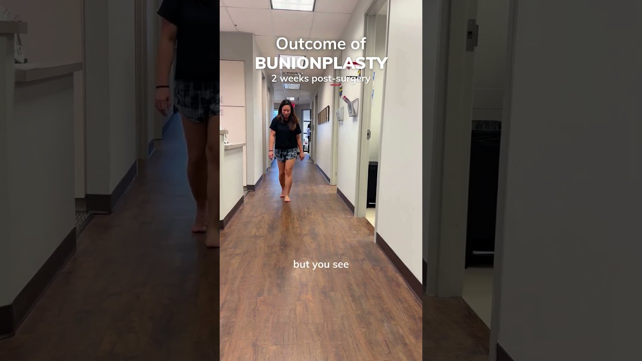
If big toe motion is limited or painful, especially with a bump at the joint, a foot and ankle arthritis specialist or a foot and ankle bunion surgeon may need to evaluate hallux rigidus or bunion mechanics. Early, conservative care can prevent a bigger problem.
Condition-specific strategies from clinic
No two ankles are the same, yet patterns emerge. The following approaches reflect what tends to work, along with when I refer to a foot and ankle surgical specialist or other colleagues.
After an ankle sprain that never felt “right” again
Chronic inversion sprains create a cycle of ligament laxity laterally, protective stiffness medially, and hesitancy from the nervous system. I address it in this order: restore dorsiflexion glide, strengthen peroneals and posterior tibialis, then rebuild balance and reactive control.
Expect 6 to 12 weeks for meaningful change. If the ankle still feels unstable, a foot and ankle ligament specialist may recommend bracing for sport or, in severe cases, consider ligament repair. A foot and ankle ligament repair surgeon typically reserves surgery for recurrent sprains with mechanical instability that fails rehab.
Achilles tendinopathy with morning stiffness
The trick is to load the tendon, not just stretch it. Eccentric calf raises are still gold, with slow 3 to 4 second lowers and a gradual increase in volume. I pair this with bent-knee soleus work, since the soleus is the workhorse in gait. If insertional pain dominates, avoid deep dorsiflexion at the bottom early on and perform raises from the floor rather than a step. A foot and ankle tendon specialist can add shockwave therapy or, rarely, injectables when symptoms linger beyond three months despite strong adherence.
Plantar fascia pain limiting first steps
Morning pain is a hallmark. Reduce it by placing a water bottle or ball next to the bed for 60 seconds of gentle rolling before your first steps. Strengthen the calf and the intrinsic foot, mobilize the big toe, and check footwear. Overly flexible shoes often provoke recurrrence. Night splints can help in stubborn cases. When conservative care fails, a foot and ankle heel pain doctor may use targeted shockwave, limited immobilization, or ultrasound-guided procedures. Surgery is rare and should be performed by a foot and ankle podiatric surgeon or foot and ankle orthopedic surgeon with experience in fascia release and careful patient selection.
Post-fracture or after immobilization
After a boot or cast, the ankle is both stiff and weak. Move systematically. Gentle joint mobilization starts first, then closed-chain dorsiflexion work, then loading. Avoid aggressive stretching in the first two weeks after immobilization ends. If hardware irritates the front of the ankle and blocks motion, a foot and ankle fracture specialist or foot and ankle trauma surgeon should assess. Hardware removal sometimes restores motion, but timing and bone healing status matter.
Arthritis that limits squat and stair descent
With arthritis, the goal shifts to symptom management and function rather than perfection on a goniometer. Keep the joint moving daily in pain-free arcs, emphasize calf strength, maintain a healthy body weight, and use rocker-bottom shoes when push-off hurts. Image-guided injections can buy time by reducing inflammation. When pain dictates life choices despite best conservative care, consult a foot and ankle arthritis doctor about surgical options. A foot and ankle reconstruction surgeon can address alignment, joint preservation procedures like cheilectomy, or joint replacement or fusion in advanced cases. The right procedure depends on age, activity demands, and joint-specific disease.
When to bring in the specialist
Self-care can go far, but certain signs deserve professional evaluation by a foot and ankle treatment specialist or foot and ankle injury doctor. Use this quick list to decide.
- Night pain that wakes you or constant rest pain. Mechanical catching, locking, or repeated giving way. Swelling that does not resolve over 2 to 3 weeks. Visible deformity, progressive bunion, or collapsing arch. Numbness, burning, or temperature changes in the foot. Diabetes, a wound that does not heal, or a history of poor circulation.
A foot and ankle healthcare provider will screen for systemic contributors, assess nerve health, and look for bone stress injuries that do not show up on early X-rays. People with diabetes benefit from a foot and ankle diabetic foot specialist who can protect skin integrity while improving mobility without causing ulcers. For wounds, a foot and ankle wound care doctor or foot and ankle wound care specialist should lead the care plan.
Tools that help and when to use them
Patients often ask whether they should buy a band, a brace, or a special insole. The answer depends on goals and stage of recovery.
- Resistance bands enable joint glides and controlled strength work at home. Green or blue bands offer useful tension for ankle pumps, inversion and eversion, and dorsiflexion mobilization. Balance pads and single-leg stands add proprioceptive training that reduces re-sprains. Start with eyes open, barefoot, and progress to narrow bases of support. Orthotics can offload the plantar fascia or stabilize an irritable midfoot, but they should not become a crutch forever. Use them alongside strength work. A foot and ankle podiatry expert can customize them if needed. Bracing supports unstable ankles during sport. Wear it when agility demands spike or terrain is uneven. Wean as strength and control return. Rocker-bottom shoes help painful push-off phases in hallux rigidus or ankle arthritis. They are a tool, not a defeat.
A foot and ankle consultant or foot and ankle clinical specialist can help you choose what adds value rather than clutter.
What a six-week plan often looks like
Week 1 to 2 focuses on pain control and restoring basic glide. Expect daily mobility work, light tissue prep, and short-foot drills. Measure dorsiflexion once a week, not daily, to avoid chasing noise.
Week 3 to 4 adds loading. Single-leg calf work, tibialis anterior raises, heel-elevated split squats. End sessions with gentle mobility moves to reinforce range.
Week 5 to 6 consolidates gains. Increase load slowly, add controlled hops only if walking and stairs are clean and pain light. Maintain two mobility sessions per week as you shift toward strength and function.
By the end of six weeks, many patients gain 3 to 6 centimeters in the knee-to-wall test and report smoother gait and easier stairs. Not everyone progresses on that curve, especially with long-standing stiffness or arthritis. If you stall for two consecutive weeks despite consistency, consult a foot and ankle medical expert or a foot and ankle orthopedic doctor to reassess.
Pediatric and adolescent considerations
Children are not small adults. A foot and ankle pediatric specialist monitors growth plates and alignment as kids return from sprains or fractures. They often recover range faster, but hypermobility can mask weakness. Build control with playful balance tasks and short foot drills rather than heavy stretching. If a child toes in, collapses their arch, or has recurring pain around growth spurts, a foot and ankle pediatric surgeon or foot and ankle gait specialist can check for torsional issues, tight heel cords, or tarsal coalitions.
The surgical safety net, used wisely
Most mobility problems resolve without the knife, but surgery has an important place. A foot and ankle corrective surgeon, foot and ankle complex surgery expert, or foot and ankle minimally invasive surgeon might be considered when:
- Recurrent ankle sprains leave you unstable despite full rehab. Bone spurs mechanically block dorsiflexion and conservative care fails. End-stage arthritis stops daily activities and nonoperative care offers no relief. Tendon tears or ruptures require repair to restore function.
A foot and ankle surgical expert will weigh options: debridement, osteophyte removal, ligament reconstruction, tendon repair, osteotomy to correct alignment, or joint replacement versus fusion. The details matter more than the label. Plan for postoperative rehab with a foot and ankle motion specialist so you do not trade pain for stiffness.
Troubleshooting common sticking points
You mobilize, you stretch, yet the ankle refuses to budge. I troubleshoot in this order:
- Reassess angle and technique. Many people let the arch collapse during knee-to-wall work, which cheats the ankle and feeds pronation without improving glide. Keep the second toe and kneecap aligned and the heel grounded. Shift emphasis from gastroc to soleus. Add seated bent-knee calf raises, which load the soleus more directly. Address the big toe. If it does not extend, your push-off suffers and the ankle stiffens as compensation. Add big toe mobility and toe flexor strength. Look up the chain. Hip stiffness or weak glutes change foot loading. Add hip external rotation and abduction work, and watch ankle motion improve. Consider pain modulation. If pain keeps motion guarded, speak with a foot and ankle pain relief doctor about short-term anti-inflammatories, topical analgesics, or targeted injections to reduce the barrier to movement.
If you still plateau, it may be time to partner with a foot and ankle specialist physician who can apply joint-specific manual therapy or image-guided interventions.
Real-world examples from clinic
A trail runner in her 40s came in after two sprains on the same ankle. Her dorsiflexion on the lunge test was 2 centimeters compared with 10 on the opposite side. We spent two weeks on talar glides, soleus-focused stretching, and peroneal strength, then added single-leg calf loading and uneven-surface balance. At week five she ran short intervals on packed dirt without a brace, pain 1 out of 10. We kept bracing for technical terrain for another month. She now maintains two mobility sessions weekly and has not re-sprained in a year.
A carpenter in his 50s with ankle arthritis could not descend stairs without a rail. Rocker-sole shoes, twice-weekly dorsiflexion mobilizations, and a bias toward soleus strength shifted his pain from 6 to 3 and gave him another 8 degrees of motion over eight weeks. He still has arthritis, but his workdays are manageable. He met with a foot and ankle arthritis doctor and decided to defer surgery while the current plan holds.
A diabetic patient with a mild plantar ulcer needed motion without shear. Working with a foot and ankle wound care specialist, we offloaded the area with a removable boot, performed gentle ankle pumps, and used non-weight-bearing calf work until the wound closed. Mobility returned without compromising skin integrity.
How to pick the right professional partner
Titles vary, so look at experience. For nonoperative care, a foot and ankle clinical specialist or foot and ankle biomechanics specialist with strong manual therapy and exercise credentials is a good starting point. For complex pain, recurrent injuries, or suspected structural problems, seek a foot and ankle orthopedic surgeon or foot and ankle podiatry surgeon who routinely treats your condition. Ask how often they perform the procedure in question and what their rehab protocol looks like. A strong team includes a foot and ankle gait specialist or foot and ankle motion specialist who will carry you through the post-op or return-to-sport phase.
A compact daily flow that fits busy schedules
Use this 10 to 12 minute routine on most days to maintain gains.
- One minute of light warm-up, then 90 seconds of calf and soleus soft tissue work. Two sets of banded ankle mobilizations, 10 reps each. One straight-knee and one bent-knee calf stretch, 30 to 45 seconds each. Two sets of 10 to 12 slow calf raises and 10 tibialis anterior raises. Eight short-foot holds and five slow big-toe roll-throughs.
If your day demands more, add a third set of calf raises or a set of heel-elevated split squats. If pain flares, scale volume before skipping the session entirely. Consistency beats heroics.
Final thoughts grounded in practice
Feet and ankles adapt, but they do not negotiate. Give them clear inputs and they respond. Most people do not need exotic equipment or endless passive treatments. They need permission to move into new ranges, a plan to strengthen there, and time. A foot and ankle medical surgeon or foot and ankle specialist surgeon is your ally if structure blocks progress, and a foot and ankle sports medicine doctor becomes invaluable when you need the ankle to perform at speed again.
If you feel stuck, measure your range, adjust your technique, and look for local expertise. The right foot and ankle care provider will help you distinguish between what must be mobilized, what must be strengthened, and what must be protected. With that clarity, regain movement becomes not just possible, but inevitable.